The fully automatic cell counter and viability analyzer is favored by biopharmaceutical companies in the fields of antibodies, vaccines, cell therapy, and gene therapy due to its automation, standardization, compliance with regulations, and high throughput. However, this type of product has long been dominated by imported brands, and there has been no mature solution for fully automatic fluorescence staining methods.
Focusing on cell analysis and biological reaction process monitoring technology, BioAces has applied live cell imaging and microfluidic chip technology to innovatively develop the CytScop®Pro series of fully automatic cell counters and viability analyzers that integrate trypan blue exclusion and AOPI dual fluorescence staining methods. This fills the gap in domestic equipment in the field of fully automatic cell counting and viability analysis.
Next, let me guide you through a two-part series to explore the innovative technology and excellent performance of CytScop®Pro, the new domestic equipment force. In this episode, we will first discuss the innovative technology of CytScop®Pro.
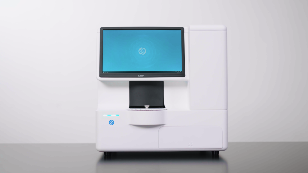
Fig.1 CytScop®Pro
Innovative Technology Chapter
01、Patented Innovative Microfluidic Chip
CytScop®Pro utilizes patented microfluidic chip technology, whose design concept is derived from the pain points that customers encounter in the traditional cell staining process, such as the crystallization of dyes, especially Trypan Blue, due to changes in storage conditions, resulting in a large amount of interfering impurities; insufficient purity of dyes; the danger of accidentally staining hands; excessive damage to cells caused by manual operation, leading to inaccurate results...
Traditional cell counting and staining requires manual mixing of dyes and samples. In the process of manual staining, it not only increases human error, but also potentially causes secondary damage to the cell samples during the mixing process, leading to deviations in the measurement of viability.
To address these pain points, BioAces has designed and developed a disposable microfluidic chip (as shown in Figure 2) that enables automatic cell staining, ushering in a new era of automated cell staining.
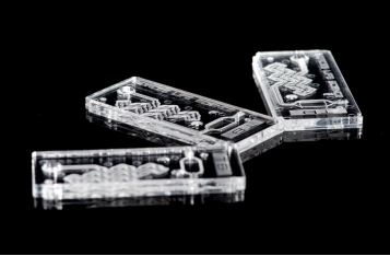
Fig.2 microfluidic chip
Innovation: Industry-leading TB Dye Embedding Technology
With a special dye formula, this technology can completely avoid dye crystallization issues, enhancing measurement efficiency. The measurement system's design is free of dye pipelines, eliminating the occurrence of pipeline blockage and significantly reducing maintenance costs.
Precision: Machine Sampling Replaces Manual Sampling
Accurate control of the dye staining ratio ensures a measurement deviation between consumables of less than 3%, reducing human error. High-purity dyes, free of impurities, minimize counting interference and enhance measurement accuracy.
Cutting-edge: Microfluidic Counting
The unique microfluidic design allows for rapid redissolution of the dye, enabling automatic cell staining. Fluid dynamics controls cell flow, ensuring the uniformity of sample concentration and enhancing measurement accuracy.
Safety: Non-contact Staining
Embedding the dye in consumables significantly reduces the risk of human exposure, ensuring safety and reliability. Waste disposal is convenient, allowing for biological neutralization after measurements are completed.
02、Streamlined Fluid Flow System
For automatic cell counters, the fluid flow system not only affects the accuracy of measurement results but also determines the sample measurement time to a certain extent. CytScop®Pro took into account the importance of fluid flow from the beginning of its product design, introducing a brand-new and streamlined fluid flow system that not only enhances mixing effect but also reduces cell damage compared to traditional fluid flow systems.
Excellent Mixing Effect
Randomly selecting 4 CytScop®Pro devices to measure cell samples with different static placement times within 0-2 hours, all of them showed good mixing effect, with a measurement concentration variation within 3% (as shown in Figure 3), indicating no significant difference.
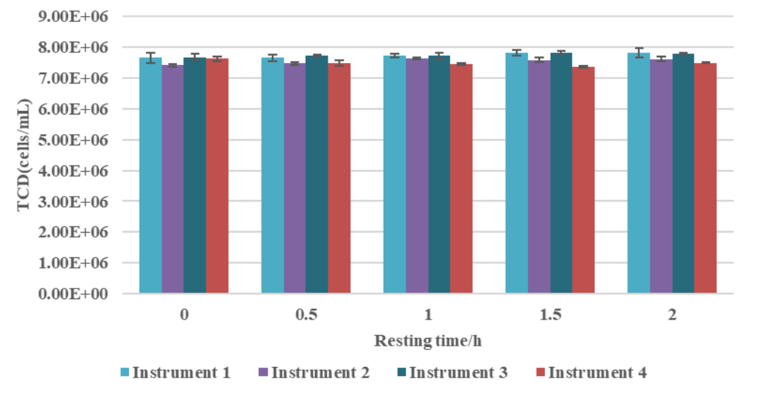
Fig. 3 Changes in total cell concentration measured by 4 instruments at different resting times
Minimal Cell Damage
To verify the damage to cells during the mixing process using CytScop®Pro, we performed viability tests on cell samples that had been left static for 0-2 hours. The results are shown in the figure below. The results indicate that CytScop®Pro's mixing system does not cause significant mechanical damage to cells, with viability changes within 5%.
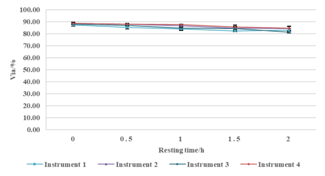
Fig. 4 Changes in cell viability measured by 4 instruments at different resting times
No Pipeline Blockage
The only component in the CytScop®Pro system that comes into contact with the fluid flow is the sample injection needle, and the flow path cleaning is also focused solely on the injection needle. This simplified fluid path design eliminates the issue of pipeline blockage.
No Contamination
Using pure water as the cleaning solution ensures no impurity contamination and reduced measurement interference.
03 Unique Optical Design
CytScop®Pro incorporates both bright-field imaging and fluorescence imaging capabilities, offering both trypan blue staining and AOPI fluorescence staining measurement modes in a single instrument. It is suitable for various cell types, enabling dual-use with significant cost savings. The optical system utilizes wide-field and large-view imaging technology, featuring the following advantages:
Two staining modes with dual fluorescence channels, suitable for multiple cell types, saving costs
Unique optical design that enhances the contrast between live and dead cells, improving analysis accuracy
Wide-field and large-view imaging provides a large statistical sample size, reducing random errors, and ensuring measurement accuracy
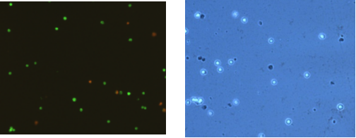
Fig. 5 AOPI staining and TB staining
04 Improved High-Precision Algorithm
Compared to traditional algorithms, the high-precision algorithm applied in CytScop®Pro is capable of accurately identifying various complex cell lines (as shown in Figure 6), demonstrating excellent compatibility and adaptability to cells in diverse environments. Additionally, the ongoing development of intelligent AI algorithms provides a solid foundation for future customized services to meet client needs.
Features include:
n Accurate identification of complex samples, suitable for aggregated cells and complex cell lines
n High scalability, allowing for adjustment of counting parameters based on the actual conditions of the sample
n Strong adaptability, ensuring better adaptability and precision while reducing the need for manual intervention
n Customization for special samples, providing tailored services to clients with specific requirements
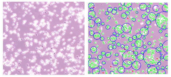
Fig. 6 Original vs. recognized image